



Introduction
The ureters are narrow tubes that carry urine from the kidneys to the bladder. When part of a ureter becomes narrowed, urine may not drain as freely as it should. This narrowing is known as a ureteric stricture, also referred to as a ureteral stricture.
A ureteric stricture can affect urine flow and may lead to symptoms such as flank pain, recurrent urinary infections or changes seen on imaging. In some cases, it may be found during investigation for another urinary concern.
Understanding what a ureteric stricture is, why it may occur and when assessment may be helpful to guide the appropriate next steps.
If you have been told you may have a ureteric stricture or you are experiencing ongoing urinary symptoms, an assessment may help determine the cause and guide appropriate management.
What Is a Ureteric Stricture?
A ureteric stricture is a narrowing of the ureter, the tube that carries urine from the kidney to the bladder.
When the ureter becomes narrowed, urine may not drain normally. This can sometimes cause urine to back up toward the kidney, which may lead to swelling of the kidney, also known as hydronephrosis.
The effect of a ureteric stricture can vary. Some patients may experience symptoms, while others may only become aware of it after imaging or investigation.
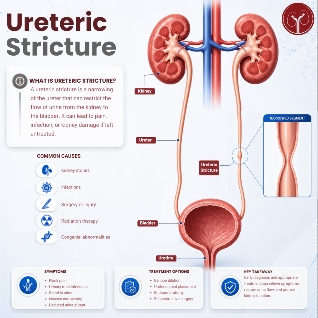
Ureteric Stricture vs Urethral Stricture
A ureteric stricture is different from a urethral stricture.
- The ureter carries urine from the kidney to the bladder.
- The urethra carries urine from the bladder out of the body.
This distinction is important because the symptoms, assessment and treatment options may differ.
Common Symptoms of Ureteric Stricture
Symptoms can vary depending on the degree of narrowing, whether urine flow is affected and whether infection is present.
Possible symptoms may include:
- Flank or side pain
- Lower abdominal discomfort
- Recurrent urinary tract infections
- Fever or chills if infection is present
- Nausea in some cases
- Blood in the urine
- Reduced kidney drainage seen on imaging
Some patients may have no obvious symptoms and the stricture may only be detected during tests for another urological concern.
What Causes a Ureteric Stricture?
A ureteric stricture may develop for different reasons. In some cases, the cause may be related to scarring, inflammation, injury or previous treatment.
Possible causes include:
Kidney stones
A stone passing through the ureter or previous treatment for kidney stones, may contribute to narrowing in some cases.
Previous surgery
Certain abdominal, pelvic or urological procedures may affect the ureter.
Congenital narrowing
Some strictures may be present from birth and only become noticeable later, depending on severity.
Inflammation or infection
Repeated inflammation may contribute to scarring and narrowing.
Radiation therapy
Radiation treatment to the abdomen or pelvis may affect nearby tissues, including the ureter.
External pressure
In some cases, narrowing or blockage may occur because of pressure from surrounding tissue or other medical conditions.
The underlying cause should be assessed carefully, as this may influence treatment planning.
When Should Ureteric Stricture Be Assessed?
Ureteric stricture should be assessed when symptoms are ongoing, recurrent or associated with possible obstruction or infection.
Assessment may be important if you experience:
- Persistent flank or side pain
- Recurrent urinary tract infections
- Fever or chills with urinary symptoms
- Blood in the urine
- Kidney swelling noted on imaging
- Ongoing symptoms after kidney stone treatment
- Unexplained changes in kidney function
Some symptoms may be mild, while others may require more urgent evaluation, especially if infection or obstruction is suspecte/space
How Is a Ureteric Stricture Diagnosed?
Diagnosis usually begins with a discussion of symptoms and medical history, followed by appropriate tests.
Assessment may include:
- A clinical history and examination
- Urine tests
- Blood tests to assess kidney function
- Ultrasound
- CT scan or CT urogram
- Specialised imaging to assess urine drainage
- Endoscopic assessment in selected cases
The aim is to understand where the narrowing is, how severe it is and whether it is affecting kidney drainage.
Treatment Options for Ureteric Stricture
Treatment depends on several factors, including the location and length of the stricture, the cause, symptoms, kidney function and whether infection or obstruction is present. Management may include measures to relieve obstruction, medicines for infection when present, or surgical options depending on the cause and severity.
Monitoring and follow-up
In selected cases, especially where symptoms are minimal and kidney drainage is not significantly affected, monitoring may be considered.
Temporary drainage
If urine flow is blocked or infection is a concern, temporary drainage may be needed. This may involve a ureteric stent or another drainage procedure depending on the clinical situation. Ureteral stents are used to help urine move from the kidney to the bladder when drainage is impaired.
Endoscopic treatment
In some cases, minimally invasive treatment may be considered. This may involve widening the narrowed area or treating scar tissue internally.
Surgical reconstruction
For longer, recurrent or more complex strictures, reconstructive surgery may be considered. The specific approach depends on the position and extent of the narrowing.
The most appropriate option can only be determined after assessment and discussion with a urologist.
What Should Patients Discuss With Their Urologist?
If a ureteric stricture is suspected or confirmed, useful questions may include:
- Where is the narrowing located?
- Is urine drainage from the kidney affected?
- What may have caused the stricture?
- Is kidney function affected?
- Is treatment required now, or can it be monitored?
- What treatment options are suitable in my case?
- Will a stent or further imaging be needed?
- What follow-up is recommended?
These questions can help patients understand the condition and the reasons behind treatment planning.
Understanding Your Next Steps
Ureteric stricture management is not the same for every patient. Some cases may require monitoring, while others may need drainage, endoscopic treatment, or reconstructive surgery.
The key step is proper assessment. This helps determine whether the narrowing is affecting urine flow, whether the kidney is under pressure, and what treatment approach may be appropriate.
A urologist can provide assessment and guidance based on your individual symptoms, imaging results, and clinical findings.
Summary
A ureteric stricture is a narrowing of the ureter, which may affect the flow of urine from the kidney to the bladder.
Symptoms may include flank pain, recurrent urinary infections, blood in the urine or kidney swelling seen on imaging. Causes may include kidney stones, previous surgery, inflammation, congenital narrowing, or other conditions affecting the ureter.
Treatment depends on the cause, location, severity and effect on kidney drainage. Assessment helps guide the most appropriate next steps
If you have been told you may have a ureteric stricture or you are experiencing ongoing flank pain, recurrent urinary infections or urinary symptoms, a consultation may help determine the cause and guide appropriate next steps.
An assessment typically involves a discussion of your symptoms and may include further evaluation if required.
Disclaimer
This article is intended for informational purposes only and does not replace professional medical advice, diagnosis, or treatment. Individual care decisions should always be made in consultation with a qualified healthcare professional.